Synthesis and Properties of Conjugates between Silver Nanoparticles and DNA-PNA Hybrids
Received 5 October 2012; Accepted 19 November 2012
Journal of Self-Assembly and Molecular Electronics, Vol. 1, 69–84.
doi: 10.13052/same2245-4551.113
Copyright © 2013 River Publishers. All rights reserved.
Gennady Eidelshtein1, Shay Halamish1, Irit Lubitz1, Marcello Anzola2, Clelia Giannini2, *, Alexander Kotlyar2, *
- 1Department of Biochemistry and Molecular Biology, George S. Wise Faculty of Life Sciences and The Center of Nanoscience and Nanotechnology, Tel Aviv University, Ramat Aviv 69978, Israel
- 2Dipartimento di Chimica, Università degli Studi di Milano, via Venezian 21, 20133 Milano Italy
- *Corresponding authors: s2shak@post.tau.ac.il; clelia.giannini@unimi.it
Abstract
We describe the preparation and properties of a stable conjugate between two 15 nm silver nanoparticles (AgNPs) and a DNA-PNA hybrid composed of 10 guanine-cytosine base pairs. We show that the conjugate is spontaneously formed during incubation of a DNA-PNA hybrid, containing phosphorothioate residues at both ends of the DNA strand with AgNPs. The conjugate molecules were separated from individual AgNPs and multiparticle structures by gel electrophoresis. We demonstrate that the absorption spectrum of the conjugate is broader than that of AgNPs, due to the interparticle plasmon coupling.
Keywords
- Silver nanoparticles
- PNA-DNA hybrid
- nanomaterials
- TEM
1. Introduction
DNA driven self-assembly of nanoparticles has proved to be useful for the synthesis of novel functional nanomaterials [1]. Relatively simple nanostructures, composed of a small number (2 to 10) of nanoparticles connected by DNA [2–7] as well as more complex two- and three-dimensional ones [8, 9] can be fabricated. In addition to unique optical properties, noble metal nanoparticles-DNA conjugates may exhibit electrically conductive or semi-conductive behavior and thus, serve as elements in nanoelectronics devices and circuits. Electronic transport measurements on double stranded (ds) DNA molecules have so far yielded very controversial results [10, 11]. Main challenges of direct conductivity measurements are associated with establishing direct physical contact between the DNA and metal electrodes and preserving the native ds conformation of the nucleic acid polymer during conductive measurement under ambient conditions. The environmental factors (e.g. pH, temperature, ionic strength of the medium) greatly affect the stability of the double stranded helix. At low ionic strength the negatively charged strands have a strong tendency to separate from each other. The preparation of samples for direct electrical measurements commonly includes deposition of DNA on electrodes followed by rinsing the surface with distilled water and drying. This treatment can thus lead to the strand separation and, as a result, to a dramatic reduction of the DNA conductivity.
In contrast to dsDNA, stability of PNA-DNA hybrids is independent of ionic strength. PNA (Peptide Nucleic Acid) is a synthetic polymer that composed of nucleic bases connected by peptide bonds [12]. The PNA-DNA hybrid adopts a ds-helical conformation that is very similar with respect to ππ interactions between nucleic bases to a native DNA [13]. PNA is uncharged at neutral pH unlike DNA, and no repulsion occurs between the complementary strands in dsPNA-DNA hybrids. The absence of interstrand repulsion governs high stability of the hybrids under a wide range of experimental conditions and makes the molecule potentially useful for nanoelectronics.
Here we report synthesis of a PNA-DNA hybrid composed of a PNA strand, (pG)10 and a complementary DNA strand, (dC)10 as well as preparation of conjugates between the hybrid and 15 nm AgNPs. Optical properties and molecular morphology of individual conjugates were measured by absorption spectroscopy and transmission electron microscopy (TEM).
2. Experimental Procedures
Unless otherwise stated, reagents were obtained from Sigma-Aldrich (USA).
2.1. DNA Oligonucleotides
Cytosine-rich oligonucleotides: [5’-(da)10-(dC)10-(da)10], composed of 10 deoxycytidine fragment (dC)10 flanked by two runs of phosphorothioated adenosines, (da)10 on either side of the strand and (dC)10, composed of 10 deoxycytidines, were purchased from Alpha DNA (Canada). These Coligonucleotides were purified on a C8 4.6 × 250 mm (Supelco Inc.) reverse-phase HPLC column. Elution was performed with a linear Methanol gradient from 0 to 50% in 30 mM K-Pi, pH 7.5 for 85 min at a flow rate of 0.7 mL/min at ambient temperature. Chromatography was performed on Agilent 1100 HPLC system. The oligonucleotides eluted from the column were desalted using a pre-packed Sephadex G-25 DNA-Grade column equilibrated with 2 mM Tris-Acetate, pH 7.5. Quantification of oligonucleotides was done by UV spectrophotometry using extinction coefficients of: 7.4, 15.4 and 11.4 mM−1 cm−1 at 260 nm for C-, A- and G-bases respectively [14]. Concentration of the 5’-(da)10-(dC)10-(da)10 was quantified, using an extinction coefficient of 382 mM−1 cm−1 at 260 nm.
2.2. PNA Oligonucleotide
A PNA strand, containing 10 G-bases, a Fluorescein moiety at N-terminus and a NH2 group at C-terminus of the sequence, Flu-(pG)10, was synthesized on MBHA resin using a 20 μmol scale Boc protocol essentially as described [15]. The synthesis was performed manually in 8 mL reaction vessel, filled with 100 mg of polystyrene resin beads. The synthetic procedure included 10 Boc deprotection-coupling-washing cycles. The synthesized G-decamer was conjugated with N-Fmoc-6-aminohexanoic (Fmoc-Ahx) pre-activated in a similar manner, and the Fluorescein isothiocyanate was subsequently attached to the N-terminus of Ahx. The kinetics of the PNA-oligomer extension and the conjugation reaction were controlled by ESI and MALDI. The product of the synthesis was purified on a 9.4 × 250 mm ZORBAX 300SBC18 reverse-phase HPLC column (Agilent Technologies, USA). The elution was in 0.1% TFA at a flow rate of 3 mL/min with a linear acetonitrile gradient from 5 to 40%. The oligonucleotide was dried by liophylization. The second purification step included ion-exchange HPLC at alkaline pH. The solubility of Flu-(pG)10 in aqueous medium at neutral pH is very low. At alkaline pH the oligonucleotide however becomes highly soluble, as a result of deprotonation of G-bases. Flu-(pG)10 was dissolved in 0.5 M LiOH and chromatographed on an anion-exchange HiTrap QHP, 5 × 1 mL FPLC column (AmershamBiosciences, USA) in 0.1 M NaOH containing 10% acetonitrile. The elution was with linear NaCl gradient from 0.5 to 1 M at a flow rate of 0.7 mL/min. The major peak fraction was collected, loaded onto a Sephadex NAP-25 DNA-Grade column, 15 × 50 mm (GE Healthcare, USA), equilibrated with 2 mM Tris-Acetate, pH 7.5 and eluted with the same buffer. The PNA solution was placed into plastic (1.5 mL capacity) tubes and stored at 4°C.
2.3. PNA-DNA Hybrid
To prepare a ds-Flu-labeled PNA-DNA hybrid, Flu-(pG)10-[(da)10-(dC)10-(da)10], HPLC purified Flu-(pG)10 and (da)10-(dC)10-(da)10 were mixed at a 2:1 molar ratio in 2 mM Tris-Acetate, pH 7.5. The mixture was heated to 80°C for 15 min and left for 2 to 3 hours at ambient temperature. NaCl was then added to a final concentration of 100 mM. The clear solution turned turbid after 30 min due to precipitation of Flu-(pG)10; the hybrid does not precipitate under these conditions. The sample was then centrifuged in a 5424 Eppendorf bench-top centrifuge at 14,000 rpm for 5 min. The pellet containing Flu(pG)10 was discarded and the supernatant, containing the ds-PNA-DNA was transferred to an Eppendorf tube and stored at 4°C. Concentration of the hybrid was quantified, using an extinction coefficient of 497 mM−1 cm−1 at 260 nm.
2.4. 15 nm AgNPs
180 mL of cooled DDW/filtered water were added into a 0.5 L glass beaker placed in an ice-water bath. 0.45 mL of 0.1 M AgNO3, 0.90 mL of 50 mM sodium citrate and 0.75 mL of 0.6 M NaBH4 were consequently added into the beaker under vigorous stirring. The yellow solution was stored at 4°C for 12–16 h. 0.72 mL of 2.5 M LiCl were then added under constant stirring at ambient temperature. The solution was transferred into 15 mL capacity DuPont Pyrex tubes and centrifuged at 14,000 rpm for 1.5 h at 20°C in a Sorval SS-34 rotor. A fluffy pellet was collected. Concentration of AgNPs was estimated spectroscopically using extinction coefficient of 2 × 10⁹ at 400 nm.
Coating of AgNPs with (dA)10, an oligonucleotide composed of 10 deoxyadenosines, was conducted as follows: 20 μM (dA)10 was added to 4 mL of AgNPs (OD∼ 90 at 400 nm). NaCl was then added to a final concentration of 25 mM and the solution was left at ambient temperature for 1 h. The concentration of the salt was increased up to 50 mM. One hour later the concentration of NaCl was adjusted to 100 mM, the solution was incubated at ambient temperature for another 80 min and loaded into a Sepharose 6B-CL column (1.6 × 35 cm). Elution was with 10 mM Na-Pi, pH 7.4 at ambient temperature. The yellow eluate was collected into 1.5 mL Eppendorf tubes and centrifuged at 13,000 rpm for 40 min at RT on bench-top centrifuge 5424 (Eppendorf, Germany). The fluffy pellet was collected and stored in dark at ambient temperature. The resulting nanoparticles were screened for their size and uniformity by TEM, revealing an average diameter of 15 ± 3 nm. The visible spectra showed a characteristic absorption peak at 400 nm. Concentration of the particles was calculated using an extinction coefficient (ε) of 2 × 109 cm−1 at 400 nm [16].
2.5. AgNP-DNA-PNA Conjugates
AgNPs were mixed with Flu-(pG)10-[(da)10-(dC)10-(da)10] at different molar ratios and incubated for 16 h in 5 mM K-Pi, pH 7.5, containing 100 mM NaCl at ambient temperature. The incubation mixture was electrophoresed on a 1.5% agarose gel. The gel areas were cut out from the gel with a razor blade and the conjugates were electroeluted into dialysis bags. Electroeluted samples were centrifuged at 13,000 rpm for 40 min on a 5424 Eppendorf bench-top centrifuge. The pellets were suspended in a small (20–100 μL) volume of TEA buffer.
2.6. Gel Electrophoresis
Samples were loaded into a 7×7 cm 1.5% agarose gel, and electrophoresed at 4°C and 130 V for 45 min, using TAE as a running buffer. The gel was stained with ethidium bromide (5 μg/mL) for 20 min. The DNA was visualized with a Bio Imaging System 202D at 302 nm.
2.7. Absorption Spectroscopy
Absorption spectra were acquired with a UV/VIS Evolution-60 spectrophotometer from Thermo Scientific (USA). Measurements were conducted at 25°C in a wavelength range from 350 to 800 nm.
2.8. HPLC
Chromatography assays were done on Agilent 1100 HPLC system, (Hewlett Packard, USA), including a photodiode array detector unit with quartz flow cells with 1 cm optical path.
2.9. TEM Spectroscopy
TEM images were acquired by using carbon-coated grids (400 mesh). 2.5 μL of a sample in 40 mM Tris-Acetate, pH 7.8, were dropped onto a grid surface. After incubation for 5 min at ambient temperature, the excess solution was removed by blotting with a filter paper. TEM imaging was performed on a TEM (JEM model 1200 EX instrument) operated at an accelerating voltage of 120 kV.
3. Results
3.1. Preparation and Characterization of a DNA-PNA Hybrid
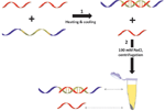
Synthesis of the hybrid includes (see Figure 1): (1) Heating a mixture of HPLC purified PNA and DNA strands (PNA to DNA molar ratio is 2) to 80°C and slow cooling the sample down to ambient temperature; (2) addition of 100 mM NaCl, incubation of the mixture at room temperature for an hour and separation of the hybrid from the excess of Flu-(pG)10 by centrifugation.
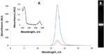
We have shown that the hybrid elutes as a single peak from a size-exclusion HPLC column (Figure 2A) and is characterized by absorption maxima at 258 and 495 nm (see inset in Figure 2A). The absorption at 490 and 260 nm reflects the relative amounts of fluorescein and nucleic bases in the hybrid respectively. The spectrum corresponds nicely with the molecule composed of 10 G-, 10 C-bases and one fluorescein residue. The hybrid migrates through the agarose gel as a single narrow band (Figure 2B). The results presented in Figure 2 thus show that the hybrid is stable and does not dissociate into single strands during HPLC and electrophoresis.
3.2. Synthesis of Oligonucleotide-Coated Silver Nanoparticles
The citrate-protected 15 nm AgNPs are unstable and precipitate out of the solution at salt concentrations higher than 30 mM. In order to increase their stability we have coated them with an oligonucleotide composed of 10 deoxyadenines, (dA)10. Incubation of AgNPs with large (300–500 fold) molar excess of each of the following sequences: (dA)10, (dC)10 or (dG)10 greatly increased the resistance of the particles to salts. The above oligonucleotide-coated particles did not precipitate at 100 mM NaCl in contrast to citrate-protected ones that aggregate at salt concentrations exceeding 30 mM. In contrast, (dT)10 was incapable of stabilizing the particles.
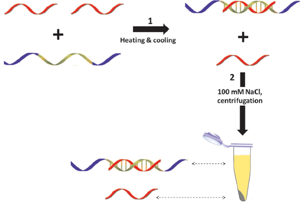
Figure 1 Schematic representation of Flu-(pG)10-[(da)10-(dC)10-(da)10] synthesis. Flu(pG)10 is depicted in red, [(da)10-(dC)10-(da)10] in green and blue; blue fragments correspond to (da)10.
We have also demonstrated that incubation with ATP did not affect the resistance of citrate-protected AgNPs to salts. The particles coated with (dA)3, (dC)3 or (dG)3 did not aggregate at 50 mM NaCl, but precipitated at higher salt concentrations (data not presented). These results clearly show that stability of nanoparticles depends on the length of oligonucleotide used for coating.
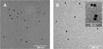
The (dA)10-coated particles can be chromatographed and electrophoresed in contrast to citrate-protected ones that do not enter columns and gels. This property enables one to use size-exclusion chromatography (see Section 2) for purification of the particles from high molecular weight aggregates that are always present in the suspension. The size-exclusion chromatography yielded monodisperse spherical nanoparticles with narrow size distribution as revealed by TEM analysis (see Figure 4A). As seen in the TEM image the average size of the particles is equal to 15 ± 3nm.
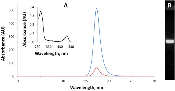
Figure 2 Size-exclusion HPLC (A) and electrophoresis (B) of Flu-(pG)10-[(da)10-(dC)10-(da)10]. The PNA-DNA hybrid was prepared as shown in Figure 1. (A) The hybrid was loaded on a size-exclusion G-4000-DNA-PW HPLC column (7.8 × 300 mm) from Toso (Japan) and was isicratically eluted in 20 mM Tris-Acetate, pH 8.0. The elution was followed at 260 (blue curve) and 495 nm (red curve). The inset shows a UV/VIS spectrum of the eluted hybrid. (B) 20 μL of the hybrid solution were loaded on a 1.5% agarose gel (7×7 cm). Electrophoresis was conducted at 4°C and 130 V for 40 min in TEA buffer. The gel was stained with ethidium bromide.
3.3. Synthesis of Conjugates between the DNA-PNA Hybrid and AgNPs
The phosphorothioated residues can covalently anchor the hybrid ends to the surface of silver and gold particles [6, 17], yielding nanoparticle-DNA-PNA conjugates. We have shown that incubation of (dA)10-coated AgNPs with the phosphorothioate-functionalized hybrid, (pG)10-[(da)10-(dC)10-(da)10] as described in Experimental Procedures section yielded structures that move through the gel slower than individual AgNPs. A new yellow band (marked by arrow “2”, see lane 3 in Figure 3) is clearly seen in the gel image. Incubation of AgNPs with the hybrid not containing phosphorotioated residues, (pG)10-(dC)10, under identical conditions did not yield the above band (see Figure 3, lane 2). Slices corresponding to bands “1” and “2” (see Figure 3) were cut out of the gel with a razor blade. The molecules were electroeluted from the slices and characterized by TEM as described in Section 2.
TEM analysis (see Figure 4) shows that the waste majority of structures electroeluted from slices marked with arrows “1” and “2” composed of 1 and 2 nanoparticles respectively. Structures electroeluted from the yellow area above band “2” (see lane 3 in Figure 4) were composed of 3–4 particles (data not presented). The interparticle separation distance in the conjugates is approximately equal to 3–4 nm (see inset in the right panel of Figure 4B) that corresponds nicely with the length of a ds-PNA-DNA linker.
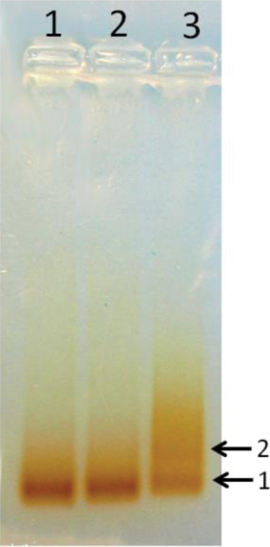
Figure 3 Electrophoretic separation of AgNPs-DNA-PNA conjugates. AgNPs (OD at 400 nm is equal to 800) (lane 1) were incubated with 0.8 μM (pG)10-(dC)10 (lane 2) and 0.8 μM (pG)10-[(da)10-(dC)10-(da)10] (lane 3) in: 5 mM K-Pi, pH 7.5 and 0.1 M NaCl at ambient temperature for 24 h. 10 μL of each sample was loaded onto a 1.5% agarose gel and electrophoresed at 4°C and 130 V for 40 min.
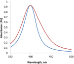
As seen in Figure 5 the absorption spectrum of the AgNP-PNA-DNA conjugate is broader than that of the particles not connected to each other. The observed broadening of Ag surface plasmon band around 400 nm reflects the coupling between closely spaced AgNPs in the structure.
We have shown that the conjugates are stable and do not dissociate into single particles at low ionic strength. Incubation for 2 days at ambient temperature in distillate water produced no effect either on the absorption spectrum or on the mobility of the conjugate band through the gel (data not shown).
4. Discussion
We have synthesized a PNA-DNA hybrid composed of a PNA strand, (pG)10 and a DNA strand functionalized with phosphorithioated adenine residues on either side of the DNA oligonucleotide, (da)10-(dC)10-(da)10. The hybrid is stable under low ionic strength conditions when the two strands of canonical DNA dissociate from each other.

Figure 4 TEM images of AgNPs and the AgNPs-PNA-DNA structures. Molecules electroeluted from gel slices corresponding to bands “1” (A) and “2” (B) (see Figure 3), were deposited on 400 mesh copper carbon grids and visualized by TEM. The inset is an enlarged image of one of the structures.
We have demonstrated that silver nanoparticles in contrast to gold ones can be stabilized by regular adenosine oligonucleotides (not containing thiols, phosphorothioates or amines). Incubation of AgNPs with a great molar excess of oligonucleotides composed of either 10 adenosine, cytosine or guanine bases, yielded stable nanoparticles that do not precipitate at salt concentrations up to 300 mM. In contrast, incubation with (dT)102 did not affect the particles stability. This result corresponds well with earlier observations, showing that thymine bases interact with the surface of silver [18] and gold [19, 20] particles weaker than other DNA bases. The week interaction of thymine thus may due to the absence of an exocyclic amine group in this base.
We have shown (see Figures 4 and 5) that incubation of (dA)10-coated AgNPs with PNA-DNA flanked with runs of ten phosphorothioated adenine-bases on both ends of the hybrid results in the formation of structures containing two nanoparticles. The absorption spectrum of the dimer is broader than that of AgNPs not connected to each other. It is well known that plasmons of closely spaced nanoparticles are strongly coupled [21, 22]. The spectrum broadening (see Figure 5) thus reflects the presence of electromagnetic coupling between plasmons of AgNPs in the dimer.
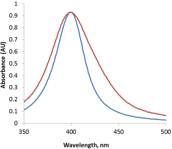
Figure 5 Normalized absorption spectra of AgNPs (blue curve) and AgNP-PNA-DNA conjugates (red curve) electroeluted from a gel slice corresponding to band “2” (see Figure 3).
The ability of DNA to conduct electrical current depends critically on π-π interaction between stacked Watson–Crick base pairs in the ds-polymer. Even partial strands separation, can strongly reduce, if not completely abolish, conductivity of the DNA molecule. The separation of DNA strands under conditions of low ionic strength might be the reason for poor conductivity reported earlier [23]. PNA-DNA hybrids lack the above disadvantage. The molecule adopts a double helical conformation that is very similar with respect to π-π interactions to a native ds-DNA even in pure water. Attachment of two 15 nm silver particles to both ends of the hybrid is also an advantage that makes possible to place the conjugate in between electrodes separated by 20–30 nm and measure conductivity of very short (∼3 nm in our case) DNA-PNA molecules. We believe that conjugates of nanoparticles with PNADNA will possess interesting conductive and optical properties leading to the development of new DNA-based nano-conductors, semiconductors and electro-optical switches.
Acknowledgments
This work was supported by the Israel Science Foundation, 172/10. M.A. acknowledges fellowship support from the Nerviano Medical Sciences.
References
[1] M. R. Jones, K. D. Osberg, R. J. Macfarlane, M. R. Langille, C. A. Mirkin Chem. Rev., 111, 3736–3827 (2011).
[2] A. P. Alivisatos, K. P. Johnsson , X. G. Peng, T. E. Wilson, C. J. Loweth, M. P. Bruchez, P. G. Schultz Nature, 382, 609-611 (1996).
[3] C. J. Loweth, W. B. Caldwell, X. G. Peng, A. P. Alivisatos , P. G. Schultz Chem. Int. Ed., 38, 1808–1812 (1999).
[4] S. Bidault , F. J. G.de Abajo, A. Polman JACS, 130, 2750–2751 (2008).
[5] A. J. Mastroianni, S. A. Claridge, A. P. Alivisatos JACS, 131, 8455–8459 (2009).
[6] I. Lubitz, A. Kotlyar Bioconjug. Chem., 22, 2043–2047 (2011).
[7] N. Borovok, E. Gillon, A. Kotlyar Bioconjug. Chem., 23, 916–922 (2012).
[8] D. Nykypanchuk, M. M. Maye, D. van der Lelie, O. Gang Nature, 451, 549–552 (2007).
[9] S. Y. Park, A. K. R. Lytton-Jean, B. Lee S. Weigand, G. C. Schatz, C. A. Mirkin Nature, 451, 553–556 (2008).
[10] D. Porath, G. Cuniberti, R. Di Felice Top Curr. Chem., 237, 183–228 (2004).
[11] R. G. Endres, D. L. Cox, R. R. P. Singh Rev. Mod. Phys., 76, 195–214 (2004).
[12] P. E. Nielsen, M. Egholm, R. H. Berg, O. Buchardt Science, 254, 1497–1500 (1991).
[13] M. Egholm, O. Buchardt, L. Christensen, C. Behrens, S. M. Freier, D. A. Driver, R. H. Berg, S. K. Kim, B. Norden, P. E. Nielsen Nature, 365, 566–568 (1993).
[14] C. R. Cantor, M. M. Warshaw, H. Shapiro Biopolymers, 9, 1059–1077 (1970).
[15] L. Christensen, R. Fitzpatrick, B. Gildea, K. H. Petersen, H. F. Hansen, T. Koch, M. Egholm, O. Buchardt, P. E. Nielsen J. Pept. Sci., 1, 175–183 (2004).
[16] J. Yguerabide, E. E. Yguerabide Anal. Biochem., 262, 137–156 (1998).
[17] I. Lubitz, A. Kotlyar Bioconjug. Chem., 22, 482–487 (2011).
[18] S. Basu, S. Jana, S. Pande, T. Pal J. Coll. Int. Sci., 321, 288–293 (2008).
[19] A. Gourishankar, S. Shukla, K. N. Ganesh, M. Sastry J. Am. Chem. Soc, 126, 13186– 13187 (2004).
[20] L. M. Demers, M. Ostblom, H. Zhang, N. H. Jang, B. Liedberg, C. A. Mirkin J. Am. Chem. Soc., 124, 11248–11249 (2002).
[21] S. K. Ghosh, T. Pal Chem. Rev., 107, 4797–4862 (2007).
[22] N. G. Khlebtsov, L. A. Dykman J. Quantit. Spectrosc. Radiative Transfer, 111, 1–35 (2010).
[23] R. G. Endres, D. L. Cox, R. R. P. Singh Rev. Mod. Phys., 76, 195–214, (2004).
Biographies

Gennady Eidelshtein received his B.Sc degree in Biotechnology from Hadassah College, Jerusalem, Israel, in 2009. In 2012 Gennady holds his Master’s degree in Materials & Nanotechnology at Tel Aviv University under supervision of Professor Alexander Kotlyar. Today Gennady is Ph.D. student in Alexander Kotlyar’s group. His research is focused on novel metal and DNA-based functional nanomaterials.

Shay Halamish was born in Jerusalem, Israel, January 1984. He recieved his B.Sc degree in Biotechnology from Bar Ilan University Israel in 2010. In 2010 Shay started his master’s degree in Materials & Nanotechnology at Tel Aviv University under supervision of Professor Alexander Kotlyar. His research is focused on optical properties of metal nanoparticles.

Irit Lubitz was born in Ramat Gan, Israel, January 1977. She received her B.Sc degree in biology from Bar Ilan University, Israel. She holds Master’s and Ph.D. degrees in Biochemistry, under supervision of Professor Alexander Kotlyar, from Tel Aviv University. Since 2005 her research has been focused on novel DNA-based functional nanomaterials.

Marcello Anzola was born in Parma, Italy, in 1986. He received his M.Sc degree in Chemistry at Università degli Studi di Parma in 2010. His research was focused on PNAs as diagnostic tools. He currently works at Nerviano Medical Sciences, an Italian research center on oncologic drugs.

Clelia Giannini graduated in Pharmaceutical Chemistry and Technology in 1996 and received her Ph.D. in Bioactive Natural Compound at University of Naples (Italy) in 2001. She joined, as post-doc, the Organic and Industrial Chemistry Department of University of Milan in 2002. She became Assistant Professor in Organic Chemistry in 2005 at the same university. Her research activity is mostly in the fields of the synthesis and structural studies of modified Peptide Nucleic Acid (PNA).

Alexander Kotlyar graduated in Biochemistry and got his Ph.D. in Biochemistry at Moscow State University in 1985. He is a faculty member of Tel Aviv University since 1994. He is an author of about 100 peer reviewed journal papers. His major research interests since 2001 focus around the DNA-based nanostructures. He developed novel methods for enzymatic synthesis of various DNA nanowires and complexes of the wires with metal particles.<page/>